VARICOSE TREATMENT
Indications for the Treatment of Varicose Veins
Indications for the treatment of superficial varicose veins are intrinsically linked to the clinical form and individual findings, with the aim of improving the patient's quality of life. Several therapeutic options are available for those with incompetent superficial veins, saphenous veins with reflux, tributary varicose veins, reticular veins and telangiectasias.
Numerous studies have demonstrated the beneficial effects of the intervention, not only in patients with chronic venous disease who exhibit skin changes, but also in those who exclusively have superficial varicose veins.
The management strategy to be adopted will depend on the specific characteristics of each patient. In summary, clinical assessment and individual factors related to the patient continue to be the basis for conducting treatment.
The first step before treating patients with varicose veins should be an ultrasound examination of the venous system. This diagnosis is essential to evaluate the superficial and deep venous system, being decisive in therapeutic planning.
Minimally Invasive Surgery
Minimally invasive alternatives should be considered for the treatment of great saphenous veins, small saphenous veins, accessory saphenous veins and other superficial veins, such as the posterior circumflex vein of the thigh (Giacomini).
The two most commonly used techniques are endovenous laser ablation and radiofrequency ablation. Other techniques may be part of the therapeutic arsenal, such as the use of foam or cyanoacrylate.
The use of thermal ablation techniques (endovenous laser ablation and radiofrequency ablation) requires the injection of tumescent fluid around the target vein. The entire procedure is performed with vascular ultrasound guidance. A laser fiber (or radiofrequency catheter) is inserted percutaneously. After successful cannulation, the catheter is advanced through an introducer sheath along the course of the vein to be treated and positioned distal to the saphenofemoral or saphenopopliteal junction.
When removing the catheter or fiber, energy is emitted intraluminally, with the intention of causing
irreversible thermal damage to the vein wall. The risk of these procedures is low. Recanalization is the most common cause of recurrence after thermal ablation, while neovascularization is more common after classical surgery.
Transdermal Laser
Transcutaneous laser uses selective photothermolysis to obliterate blood vessels while sparing surrounding tissues. When considering laser treatment for reticular veins or telangiectasias, the parameters in utilizing this method will be based on the size of the target vessel.

SOLON V Transdermal Laser
Sclerotherapy
Sclerotherapy refers to the administration of a sclerosing agent into a specific vein, with the aim of causing damage to its wall and promoting lasting fibrosis. Sclerosing agents can be used in foam or liquid form.
The foam sclerotherapy technique is preferred in anatomical scenarios that make intravenous cannulation or advancement of ablation devices challenging, and is also considered appropriate for the treatment of recurrent tortuous varicose veins.
Liquid sclerotherapy is predominantly reserved for the treatment of reticular veins and telangiectasias. It is important to highlight that the concentration of the sclerosing agent used will depend on the size of the target vein.
The majority of adverse events observed after sclerotherapy of reticular veins and telangiectasias are mild in nature, with the most frequent being transient hyperpigmentation, neovascularization and scar formation at the injection site.
CLaCS (Cryo-Laser and Cryo-Sclerotherapy)
CLaCS is the acronym for a technique that is characterized by the synergism of the transdermal laser, which uses pulses of light to heat and occlude the vessels, with sclerotherapy, consisting of the injection of a solution into the vessels.
In this procedure, it is possible to use augmented reality devices, use a phleboscope and apply cooling to the skin, aiming to improve both the effectiveness and safety of this treatment.

Transdermal laser treatment and Augmented Reality by Vein Finder
Conventional Surgery (Phlebectomy)
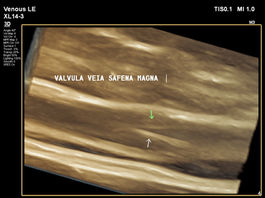
The phlebectomy technique consists of making multiple small incisions adjacent to the varicose vein, after appropriate marking in the preoperative period. Varicose vein removal is carried out using delicate hooks and small, fine-tipped surgical forceps. Often, after extrusion of the diseased vein, it is not necessary to suture the skin. When venous echodoppler shows reflux in the saphenous veins, conventional surgery remains a treatment alternative. Valvular reflux of the saphenous vein is an alteration in which the valves present dysfunctions, compromising adequate blood transport.
Conventional surgery involves larger incisions, followed by the passage of a phleboextractor, which pulls the saphenous vein under the skin. The purpose of the technique is to remove the saphenous vein segment with reflux, although this is not always feasible. Anatomical limitations and surgical technique, such as the need to only pull the saphenous vein through segmental incisions, can present challenges.
Factors such as previous surgical manipulation, tortuosity of the great saphenous vein, small diameter saphenous vein and/or segmental competence (venous valves functioning in some segments of the saphenous vein, but not in others) and technical difficulties, including anatomical variations, may contraindicate, hinder or make it impossible to correct saphenous vein reflux by removing it.
It is worth noting that reflux is just one element in the venous echodoppler examination and should be valued when associated with the assessment of venous diameter, clinical data and specific symptoms.
.


